Home » Without Label » Upper Leg Tendon Anatomy : Anatomy Physiology 1 Sayers Flashcards Ch 10 11 Muscle Tissue Studyblue Anatomy And Physiology Anatomy Leg Muscles Anatomy : When tendons become inflamed, irritated or suffer microscopic tears, the condition is called tendonitis.
Upper Leg Tendon Anatomy : Anatomy Physiology 1 Sayers Flashcards Ch 10 11 Muscle Tissue Studyblue Anatomy And Physiology Anatomy Leg Muscles Anatomy : When tendons become inflamed, irritated or suffer microscopic tears, the condition is called tendonitis.
Upper Leg Tendon Anatomy : Anatomy Physiology 1 Sayers Flashcards Ch 10 11 Muscle Tissue Studyblue Anatomy And Physiology Anatomy Leg Muscles Anatomy : When tendons become inflamed, irritated or suffer microscopic tears, the condition is called tendonitis.. This is why you have to indicate which biceps you are taking about when discussing one or other of these muscles. The anterior, or front upper leg muscles are the quadriceps. Learn its anatomy and function now at kenhub! Another large hip flexor is the rectus femoris. This important tendon in the back of the calf and ankle stores the elastic energy needed for running, jumping, and other physical activity.
This mri wrist coronal cross sectional anatomy tool is absolutely free to use. The muscles of the anterior thigh consist of the quadriceps (or quads): •medial thigh muscles•adductor longus muscle•adductor. It is the junction of the thigh and the leg and is a hinge joint. Squeeze your knees together and boom, you're contracting the adductors.
18 Tips For Bulletproof Knees T Nation Upper Leg Muscles Muscle Diagram Leg Muscles from i.pinimg.com The calf muscle, on the back of the lower leg, is actually made up of two muscles: The medial, or towards the middle of the body, upper leg. They consist of the rectus femoris, vastus intermedius, vastus lateralis and the vastus medialis. This tendon helps your leg bend when you raise your knee. It runs straight down the leg and attaches to the patella via the quadriceps femoris tendon. Upper leg anatomy and function. •medial thigh muscles•adductor longus muscle•adductor magnus muscle•adductor. The knee joint is commonly injured, so understanding its anatomy can help you understand the conditions that cause problems, so you stay safe and prepared.
When tendons become inflamed, irritated or suffer microscopic tears, the condition is called tendonitis.
17.03.2021 · upper leg tendon anatomy : The quads make up about 70% of the thigh's muscle mass. These muscles start at the bottom of your pelvis extending down the back of your thigh and along either side of your knee, to your lower leg bones. Learn its anatomy and function now at kenhub! The calf muscle, on the back of the lower leg, is actually made up of two muscles: Upper leg anatomy and function. Ebraheim's educational animated video describes muscle anatomy of the thigh. The only muscle of the quadriceps to cross both the hip and knee joints. One of the most important tendons in terms of mobility of the leg is the achilles tendon. Squeeze your knees together and boom, you're contracting the adductors. •medial thigh muscles•adductor longus muscle•adductor magnus muscle•adductor. The four muscles all extend the lower leg. The rectus femoris is one of the quadriceps muscles, the largest group of muscles on the front of the thigh.
This important tendon in the back of the calf and ankle stores the elastic energy needed for running, jumping, and other physical activity. It is also visible on the medial edge of the thigh from the anterior. The calf muscle, on the back of the lower leg, is actually made up of two muscles: Lie prone on a hamstring curl machine. The tendons for these muscles begin at your ischial tuberosity, or ischium (the bony bump under each buttock), and attach on the outer edges of your shinbones (tibia and fibula) just below the back of your knee.
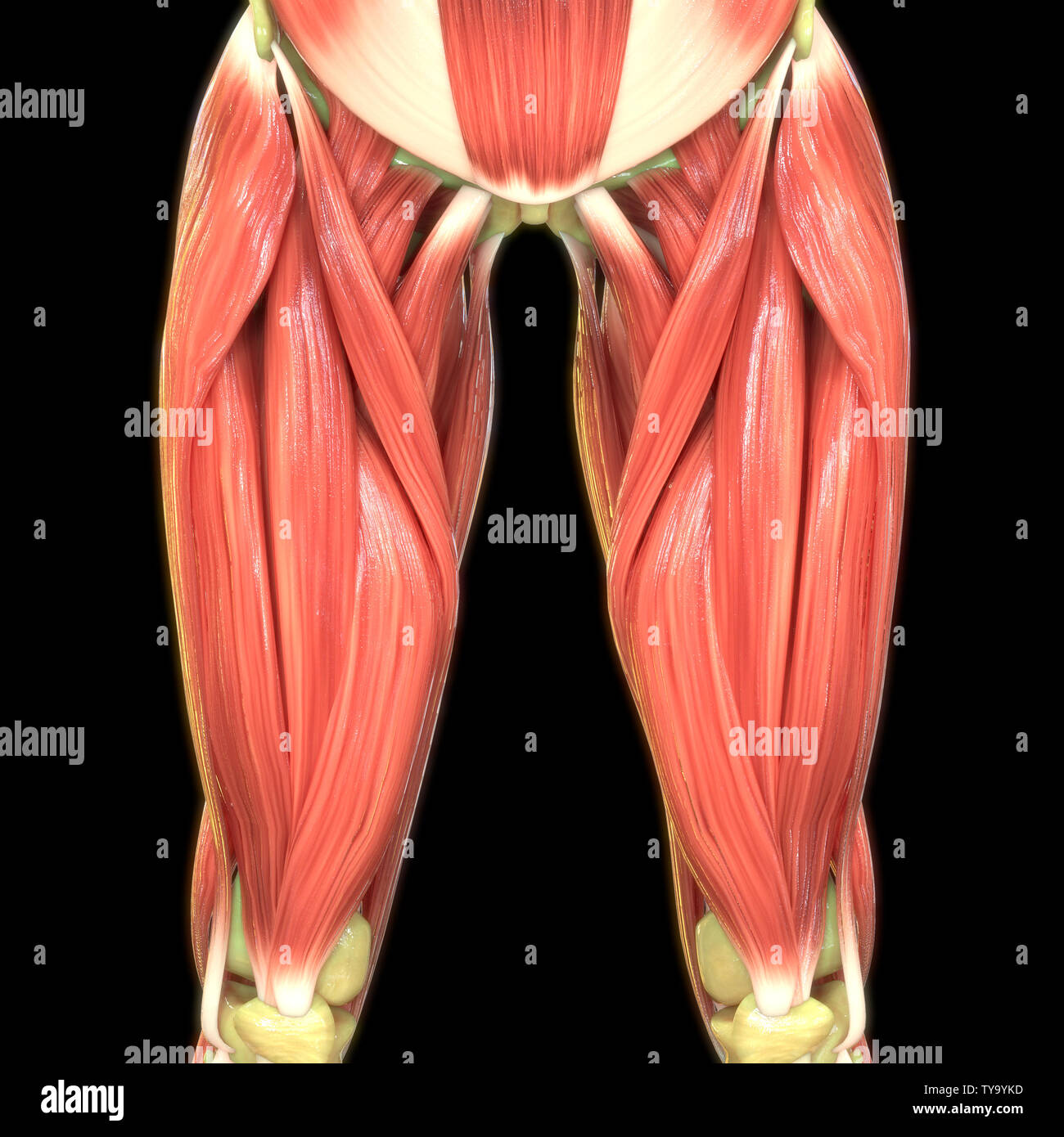
Human Body Muscles Anatomy Stock Photo Alamy from c8.alamy.com Tendons are cords made of tough tissue, and they work as special connector pieces between bone and muscle. A muscle strain (muscle pull or tear) is a common injury, particularly among people who participate in sports. Other muscles of the anterior (front) thigh include the pectineus, sartorius,. It runs straight down the leg and attaches to the patella via the quadriceps femoris tendon. Rectus femoris these four muscles come together to form a single tendon, which inserts into the patella, or kneecap. This important tendon in the back of the calf and ankle connects the plantaris, gastrocnemius, and soleus muscles to. Upper leg muscles ligaments c 1900 antique anatomy print etsy. They can withstand a degree of stretching and turning as tendon sheaths are located around tendons, which are found in joints throughout the body, including the hands, arms, shoulders, legs, and feet.the human leg, in the general word sense, is the entire lower limb of the human body.
Upper leg anatomy and function.
It arises by a thin aponeurosis from the anterior margins of the lower half of the symphysis pubis and the upper half of the pubic arch. This deep muscle begins in the low back and pelvis and connects on the inside edge of the upper femur. Upper leg anatomy and function the upper leg is often called the thigh. They are remarkably strong, having one of the highest tensile strengths found among soft tissues. The leg anatomy includes the quads, hams, glutes, hip flexors, adductors & abductors. Tendons are thick bands of tissue that connect muscles to bone. 3d anatomy tutorial on the muscles of the thigh and the gluteal region from anatomyzone for more videos, 3d models. The hamstring portion of the adductor magnus has a similar action to these muscles, but is located in the medial thigh. This mri wrist coronal cross sectional anatomy tool is absolutely free to use. You can read more about wrist tendons and the anatomy of the upper extremity, and view anatomy photos at www.handcare.org. The tendons for these muscles begin at your ischial tuberosity, or ischium (the bony bump under each buttock), and attach on the outer edges of your shinbones (tibia and fibula) just below the back of your knee. Possibly the most important tendon in terms of mobility is the achilles tendon. The medial, or towards the middle of the body, upper leg.
The tendons for these muscles begin at your ischial tuberosity, or ischium (the bony bump under each buttock), and attach on the outer edges of your shinbones (tibia and fibula) just below the back of your knee. For a more detailed anatomy of the muscle, check out the following leg muscle diagrams posted below. On the medial edge of the posterior thigh is the gracilis muscle. It runs straight down the leg and attaches to the patella via the quadriceps femoris tendon. •medial thigh muscles•adductor longus muscle•adductor.
Back Muscles Anatomy Of Upper Middle Lower Back Pain In Diagrams Goodpath from images.ctfassets.net This is why you have to indicate which biceps you are taking about when discussing one or other of these muscles. These muscles start at the bottom of your pelvis extending down the back of your thigh and along either side of your knee, to your lower leg bones. Upper leg tendon anatomy : Another large hip flexor is the rectus femoris. It serves to attach the plantaris, gastrocnemius (calf) and soleus muscles to the calcaneus (heel) bone. Like the biceps brachii in the arm, the biceps femoris muscle has two heads. The hamstrings are three muscles at the back of the thigh that affect hip and knee movement. The hamstrings refer to 3 long posterior leg muscles, the biceps femoris, semitendinosus, and semimembranosus.
The fibers run vertically downward, and end in a rounded tendon, which passes behind the medial condyle.
17.03.2021 · upper leg tendon anatomy : It flexes the thigh at the hip joint, and extends at the knee joint. •medial thigh muscles•adductor longus muscle•adductor. This tendon helps your leg bend when you raise your knee. This is why you have to indicate which biceps you are taking about when discussing one or other of these muscles. Tendons are cords made of tough tissue, and they work as special connector pieces between bone and muscle. The largest muscle masses in the leg are present in the thigh and the calf. The hamstring muscles in the back of the thigh, the quadriceps muscles in the front, and the adductor muscles on the inside. Lie prone on a hamstring curl machine. It also arises from the base of the greater trochanter and the linea aspera, the supracondylar ridge, and the lateral intermuscular septum. Other muscles of the anterior (front) thigh include the pectineus, sartorius,. It serves to attach the plantaris, gastrocnemius (calf) and soleus muscles to the calcaneus (heel) bone. Therefore the most superficial muscle of the dorsal aspect of.